Featuring 96 sharp, new images obtained with state-of-the-art technology, the Second Edition of this popular pocket atlas is a quick, handy guide to interpreting cranial magnetic resonance images. It shows readers how to recognize normal anatomic structures on MRI scans...and distinguish these structures from artifacts. Each page presents a high-resolution image, with anatomic landmarks clearly labeled. Directly above the image are a key to the labels and a thumbnail illustration that orients the reader to the location and ...
Read More
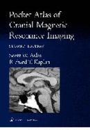
Featuring 96 sharp, new images obtained with state-of-the-art technology, the Second Edition of this popular pocket atlas is a quick, handy guide to interpreting cranial magnetic resonance images. It shows readers how to recognize normal anatomic structures on MRI scans...and distinguish these structures from artifacts. Each page presents a high-resolution image, with anatomic landmarks clearly labeled. Directly above the image are a key to the labels and a thumbnail illustration that orients the reader to the location and plane of view (sagittal, axial, or coronal). This format--sharp images, orienting thumbnails, and clear keys--enables readers to identify features with unprecedented speed and accuracy.
Read Less
Add this copy of Pocket Atlas of Cranial Magnetic Resonance Imaging to cart. $158.59, new condition, Sold by Just one more Chapter rated 5.0 out of 5 stars, ships from Miramar, FL, UNITED STATES, published 2001 by Lippincott Williams & Wilkins.